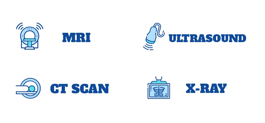
Radiology Services
At Star Imaging, we are committed to delivering advanced and accurate imaging for proper care.

Radiology Services
At Star Imaging, we are committed to delivering advanced and accurate imaging for proper care.

X-ray Services
What is an X-ray?
X-ray is an imaging study that takes pictures of bones and soft tissues. X-rays use safe amounts of radiation to create these pictures. The images help healthcare providers diagnose a wide range of conditions and plan treatments.
Most often, providers use X-rays to look for fractures (broken bones). X-ray images can help providers diagnose a wide range of injuries, disorders and diseases. X-rays are a safe and effective way for providers to evaluate your health.
Who might need an X-ray?
People of all ages, including babies, can get an X-ray. If there’s a chance you might be pregnant, tell your provider before getting an X-ray. Radiation from an X-ray can harm your fetus.
Your provider may order an X-ray to:
✔ Check for a broken bone (fracture).
✔ Identify the cause of symptoms, such as pain and swelling.
✔ Look for foreign objects in your body.
✔ Look for structural problems in your bones, joints or soft tissues.
✔ Plan and evaluate treatments.
✔ Provide routine screenings for cancer and other diseases.
How does an X-ray works?
An X-ray sends beams of radiation through your body. Radiation beams are invisible, and you can’t feel them. The beams pass through your body and create an image on an X-ray detector nearby.
As the beams go through your body, bones, soft tissues and other structures absorb radiation in different ways. Solid or dense objects (such as bones) absorb radiation easily, so they appear bright white on the image. Soft tissues (such as organs) don’t absorb radiation as easily, so they appear in shades of gray on the X-ray.
How do I prepare for an X-ray?
Tell your healthcare provider about your health history, allergies and any medications you’re taking. If you’re pregnant, think you might be pregnant or are breastfeeding (chestfeeding), tell your provider before getting an X-ray.
You usually don’t need to do anything to prepare for a bone X-ray. For other types of X-ray, your provider may ask you to:
✔ Avoid using lotions, creams or perfume.
✔ Remove metal objects like jewelry, hairpins or hearing aids.
✔ Stop eating or drinking several hours beforehand (for GI X-rays).
✔ Wear comfortable clothing or change into a gown before the X-ray.
What should I expect during an X-ray?
Depending on the type of X-ray, your provider will ask you to sit, stand or lie down on a table.
During the X-ray, your provider may move your body or limbs in different positions and ask you to hold still. You may need to hold your breath for a few seconds so the images aren’t blurry.
Sometimes children can’t stay still long enough to produce clear images. Your child’s provider may recommend using a restraint during an X-ray. The restraint (or immobilizer) helps your child stay still and reduces the need for retakes. The restraints don’t hurt and won’t harm your child.
What are the risks of an X-ray?
Although X-rays use radiation (which can cause cancer and other health problems), there is a low risk of overexposure to radiation during an X-ray. Some X-rays use higher doses of radiation than others. Generally, X-rays are safe and effective for people of all ages.
Radiation from an X-ray can harm your fetus. If you’re pregnant, your provider may choose another imaging study, such as MRI (magnetic resonance imaging) or ultrasound.
Before an X-ray, be sure to tell your provider if you might be pregnant. X-rays are a safe, effective tool providers use to help you feel better and stay healthy.
When should I know my results?
Results from a bone X-ray are usually ready within a few hours. Your provider may share your results with you after the X-ray. Results from other types of X-rays (such as a GI test) may take longer. Talk to your provider about when you can expect results.
MRI Services
What is an MRI?
An MRI (magnetic resonance imaging) scan is a painless test that produces very clear images of the organs and structures inside your body. MRI uses a large magnet, radio waves and a computer to produce these detailed images. It doesn’t use X-rays (radiation).
Because MRI doesn’t use X-rays or other radiation, it’s the imaging test of choice when people will need frequent imaging for diagnosis or treatment monitoring, especially of their brain.
What does an MRI show?
Magnetic resonance imaging (MRI) produces detailed images of the inside of your body. Healthcare providers can “look at” and evaluate several different structures inside your body using MRI, including:
✔ Your brain and surrounding nerve tissue.
✔ Organs in your chest and abdomen, including your heart, liver, biliary tract, kidneys, spleen, bowel, pancreas and adrenal glands.
✔ Breast tissue.
✔ Your spine and spinal cord.
✔ Pelvic organs, including your bladder and reproductive organs (uterus and ovaries in people assigned female at birth and the prostate gland in people assigned male at birth).
✔ Blood vessels.
✔ Lymph nodes.
When would I need an MRI?
Healthcare providers use magnetic resonance imaging (MRI) to help diagnose or monitor the treatment for many different conditions. There are also different types of MRIs based on which area of your body your provider wants to examine.
Is an MRI safe?
An MRI scan is generally safe and poses almost no risk to the average person when appropriate safety guidelines are followed.
The strong magnetic field the MRI machines emit is not harmful to you, but it may cause implanted medical devices to malfunction or distort the images.
There’s a very slight risk of an allergic reaction if your MRI requires the use of contrast material. These reactions are usually mild and controllable by medication.
Healthcare providers generally don’t perform gadolinium contrast-enhanced MRIs on pregnant people due to unknown risks to the developing baby unless it’s absolutely necessary.
Who shouldn't get an MRI?
In most cases, an MRI exam is safe for people with metal implants, except for a few types. Unless the device you have is certified as MRI safe, you might not be able to have an MRI. These devices may include:
✔ Metallic joint prostheses.
✔ Some cochlear implants.
✔ Some types of clips used for brain aneurysms.
✔ Some types of metal coils placed within blood vessels.
✔ Some older cardiac defibrillators and pacemakers.
✔ Vagus nerve stimulators.
If your healthcare provider recommends an MRI scan, they’ll ask detailed questions about your medical history and any medical devices or implants you may have in or on your body.
Who performs an MRI?
A radiologist or a radiology technologist will perform your MRI. A radiologist is a medical doctor who performs and interprets imaging tests to diagnose conditions. A radiology technologist is a highly skilled and certified healthcare professional who specializes in operating MRI scanners to produce diagnostic images.
How does an MRI work?
Magnetic resonance imaging (MRI) works by passing an electric current through coiled wires to create a temporary magnetic field in your body. A transmitter/receiver in the machine then sends and receives radio waves. The computer then uses these signals to make digital images of the scanned area of your body.
How do I prepare for an MRI?
The magnetic resonance imaging (MRI) scanner uses strong magnets and radio wave signals that can cause heating or possible movement of some metal objects in your body. This could result in health and safety issues. It could also cause some implanted electronic medical devices to malfunction.
If you have metal-containing objects or implanted medical devices in your body, your healthcare provider needs to know about them before your MRI scan. Certain implanted objects may require additional scheduling arrangements and special instructions. Other items don’t require special instructions but may require an X-ray to check on the exact location of the object before your exam.
Please tell your provider and MRI technologist if you have any of the following:
✔ Heart pacemaker or defibrillator.
✔ Electronic or implanted stimulators or devices, including deep brain stimulators, vagus nerve stimulators, bladder stimulators, spine stimulators, neurostimulators and implanted electrodes or wires.
✔ Metallic joint prostheses.
✔ Cochlear implant or other ear implants.Implanted drug pumps, such as those that pump narcotic/pain medications or drugs to treat spasticity.
✔ Programmable shunt.
✔ Aneurysm clips and coils.
✔ Stents not located in your heart.
✔ Filters, such as blood clot filters.
✔ Metal fragments in your body or eye, such as bullets, shrapnel, metal pieces or shavings.
You won’t be able to wear the following devices during your MRI. Please coordinate your MRI appointment with the day you need to change your patch or device.
✔ Continuous glucose monitor (CGM).
✔ Insulin pump.
✔ Medication patches.
In addition, tell your provider if you:
✔ Are pregnant.
✔Are not able to lie on your back for 30 to 60 minutes.
✔ Have claustrophobia (fear of enclosed or narrow spaces).
Leave all jewelry and other accessories at home or remove them before your MRI scan. Metal and electronic items aren’t allowed in the exam room because they can interfere with the magnetic field of the MRI unit, cause burns or become harmful projectiles. These items include:
✔ Jewelry, watches, credit cards and hearing aids — all of which can be damaged.
✔ Pins, metal hair accessories, underwire bras and metal zippers, which can distort MRI images.
✔ Removable dental work, such as dentures.
✔ Pens, pocket knives and eyeglasses.
✔ Body piercings.
✔ Cell phones, electronic watches and tracking devices.
How long does an MRI scan take?
The duration of an MRI scan can vary, but it typically lasts from 15 to 90 minutes, depending on the size of the area being scanned and the number of images needed.
What should I expect during an MRI?
Most MRI exams are painless, but some people find it uncomfortable to remain still for 30 minutes or longer. Others may experience anxiety from the closed-in space while in the MRI machine. The machine can also be noisy.
The general steps of an MRI scan and what to expect include:
✔ You’ll change into a hospital gown for the MRI scan.
✔ You’ll lie face up for most exams on the MRI scanning bed. The MRI scanning bed will slide into the MRI machine.
✔ As the MRI scan begins, you’ll hear the equipment making a variety of loud knocking and clicking sounds while it’s taking the images. Each series of sounds may last for several minutes. You’ll be given earplugs or headphones to wear to protect your hearing before the procedure begins.
✔ It’s important to be very still during the exam to ensure the best quality of images.
✔ It’s normal for the area of your body being imaged to feel slightly warm. If it bothers you, tell the radiologist or technologist.
✔ The MRI technologist will be able to see you and can talk with you at all times. An intercom system allows two-way communication while you’re inside the scanner. You’ll also have a call button in your hand that you can push to let the technologist know if you’re having any problems or concerns.
In some cases, your MRI may require contrast. If this applies to you, a provider will give you an IV injection of contrast material before you undergo the MRI. The IV needle may cause some discomfort but this won’t last long. You may have some bruising afterward. Some people experience a temporary metallic taste in their mouth after the contrast injection.
If you have claustrophobia, your provider may recommend a sedative drug so you feel more relaxed during the exam or even anesthesia.
What should I expect after an MRI?
If you didn’t have a sedative drug for the MRI scan, no recovery period is necessary. You can go home and resume your normal activities. If you had sedative drugs for the exam, you’ll need to recover from the effects of them before you can go home. You may need to have someone else drive you home.
When should I know the results?
After your MRI scan, a radiologist will analyze the images. The radiologist will send a signed report to your primary healthcare provider, who will share the results with you. You may need a follow-up exam. If so, your provider will explain why.
Ultrasound Services
What is an Ultrasound?
Ultrasound is a noninvasive imaging test that shows structures inside your body using high-intensity sound waves. Healthcare providers use ultrasound exams for several purposes, including during pregnancy, for diagnosing conditions and for image guidance during certain procedures.
How does an Ultrasound show?
During an ultrasound, a healthcare provider passes a device called a transducer or probe over an area of your body or inside a body opening. The provider applies a thin layer of gel to your skin so that the ultrasound waves are transmitted from the transducer through the gel and into your body.
The probe converts electrical current into high-frequency sound waves and sends the waves into your body’s tissue. You can’t hear the sound waves.
Sound waves bounce off structures inside your body and back to the probe, which converts the waves into electrical signals. A computer then converts the pattern of electrical signals into real-time images or videos, which are displayed on a computer screen nearby.
How do I prepare for an Ultrasound?
The preparations will depend on the type of ultrasound you’re having. Some types of ultrasounds require no preparation at all.
For ultrasounds of the pelvis, including ultrasounds during pregnancy, of the female reproductive system and of the urinary system, you may need to fill up your bladder by drinking water before the test.
For ultrasounds of the abdomen, you may need to adjust your diet or fast (not eat or drink anything except water) for several hours before your test.
In any case, your healthcare provider will let you know if you need to do anything special to prepare for your ultrasound.
What happens during an Ultrasound?
Preparation for an ultrasound varies depending on what body part you’ll have scanned. Your provider may ask you to remove certain pieces of clothes or change into a hospital gown.
Ultrasounds that involve applying the transducer (probe) over your skin (not in your body), follow these general steps:
✔ You’ll lie on your side or back on a comfortable table.
✔ The ultrasound technician will apply a small amount of water-soluble gel on your skin over the area to be examined. This gel doesn’t harm your skin or stain your clothes.
✔ The technician will move a handheld transducer or probe over the gel to get images inside your body.
✔ The technician may ask you to be very still or to hold your breath for a few seconds to create clearer pictures.
✔ Once the technician has gotten enough images, they’ll wipe off any remaining gel on your skin and you’ll be done.
An ultrasound test usually takes 30 minutes to an hour. If you have any questions about your specific type of ultrasound, ask your healthcare provider.
When should I know the results?
The time it takes to get your results depends on the type of ultrasound you get. In some cases, such as prenatal ultrasound, your provider may analyze the images and provide results during the test.
In other cases, a radiologist, a healthcare provider trained to supervise and interpret radiology exams, will analyze the images and then send the report to the provider who requested the exam. Your provider will then share the results with you or they may be available in your electronic medical record (if you have an account set up) before your provider reviews the results.
CT Scan Services
What is a CT Scan?
A CT (computed tomography) scan is a type of imaging test. Like an X-ray, it shows structures inside your body. But instead of creating a flat, 2D image, a CT scan takes dozens to hundreds of images of your body. To get these images, a CT machine takes X-ray pictures as it revolves around you.
Healthcare providers use CT scans to see things that regular X-rays can’t show. For example, body structures overlap on regular X-rays and many things aren’t visible. A CT shows the details of each of your organs for a clearer and more precise view.
Another term for CT scan is CAT scan. CT stands for “computed tomography,” while CAT stands for “computed axial tomography.” But these two terms describe the same imaging test.
How do I prepare for a CT Scan?
Your healthcare provider will tell you everything you need to know about CT scan preparation. Here are some general guidelines:
✔ Plan to arrive early. Your provider will tell you when to come to your appointment.
✔ Don’t eat for four hours before your CT scan.
✔ Drink only clear liquids (like water, juice or tea) in the two hours leading up to your appointment.
✔ Wear comfortable clothes and remove any metal jewelry or clothing. Your provider may give you a hospital gown to wear.
Your provider might use a contrast material to highlight certain areas of your body on the scan. For a CT scan with contrast, your provider will place an IV (intravenous line) and inject a contrast (or dye) into your vein. They may also give you a substance to drink (like a barium swallow) to highlight your intestines. Both improve the visibility of specific tissues, organs or blood vessels and help healthcare providers diagnose several medical conditions. IV contrast agents usually flush from your system (when you pee) within 24 hours.
Here are some additional preparation guidelines for a CT scan with contrast:
✔ Blood test: You might need a blood test before your scheduled CT scan. This will help your healthcare provider ensure the contrast material is safe to use.
✔ Diet restrictions: You’ll need to watch what you eat and drink for the four hours before your CT scan. Consuming only clear liquids helps prevent nausea when you receive the contrast. You can have broth, tea or black coffee, strained fruit juices, plain gelatin and clear soft drinks (like ginger ale).
✔ Allergy medication: If you’re allergic to the contrast agent used for CT (which contains iodine), you may need to take steroid and antihistamine medications the night before and the morning of your procedure. Be sure to check with your healthcare provider and have them order these medications for you if needed. (Contrast agents for MRI and CT are different. Being allergic to one doesn’t mean you’re allergic to the other.)
✔ Preparation solution: You should drink the oral contrast solution exactly as directed.
What should I expect during a CT Scan?
During the test, you’ll usually lie on your back on a table (like a bed). If your test requires it, a healthcare provider may inject the contrast dye intravenously (into your vein). This dye can make you feel flushed or give you a metallic taste in your mouth.
When the scan begins:
✔ The bed slowly moves into the doughnut-shaped scanner. At this point, you’ll need to stay as still as possible because movement can blur the images.
✔ You may also be asked to hold your breath for a short period of time, usually fewer than 15 to 20 seconds.
✔ The scanner takes pictures of the area your healthcare provider needs to see. Unlike an MRI scan (magnetic resonance imaging scan), a CT scan is silent.
✔ When the exam is over, the table moves back out of the scanner.
When should I know the results?
It usually takes about 24 to 48 hours to get the results of your CT scan. A radiologist (a physician who specializes in reading and interpreting CT scans and other radiologic exams) will review your scan and prepare a report that explains the findings. In an emergency setting, like a hospital or emergency room, healthcare providers often receive results within an hour.
Once a radiologist and your healthcare provider have reviewed the results, you’ll either have another appointment or receive a call. Your healthcare provider will discuss the results with you.

Why choose us?
At Star Imaging, with 25+ years of combined experience we're dedicated to providing high-quality outpatient diagnostic imaging services in a comfortable and caring environment. Our state-of-the-art facility is equipped with the latest technology, and our team of experienced radiologists and technologists are committed to delivering accurate and timely diagnosis.

Star Imaging
Phone: (832)391-6173
Fax: (832)391-6178
Email: [email protected]
Web: stardiagnosticimaging.com
Address: 1718 N. Fry Rd, Suite 350,
Houston TX 77084
